Centre of Molecular Structure

Centre of Molecular Structure

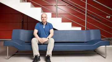
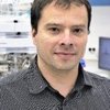
Ing. Jan Dohnálek, Ph.D.

Ing. Jan Dohnálek, Ph.D. — Laboratory head

The Institute of Biotechnology of the Czech Academy of Sciences

About us

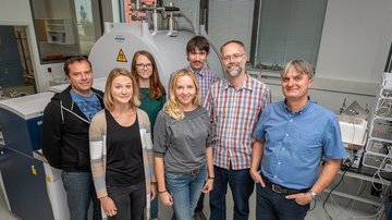
The Center of Molecular Structure (CMS) brings together several laboratories providing a comprehensive approach to the study of the spatial structure, function and biophysical properties of biological molecules. Together with the central laboratories of the CEITEC center, CMS is part of the Czech Infrastructure for Integrative Structural Biology (CIISB) - an associated national center of the European Strategy Forum on Research Infrastructures Instruct (ESFRI). CMS is also part of European infrastructure for molecular biophysics MOSBRI. CMS offers Open Access services for internal users from IBT, for other academic staff and for customers in the field of industry. It also trains Czech and foreign students and young researchers in the form of various workshops throughout the year.

CF Biophysical Techniques

The core facility Biophysical techniques enables assessment of the quality, stability and interaction properties of hundreds of biomolecular samples of many structural biology projects, be it regular checks, thorough analysis of properties or optimization of molecular constructs or handling protocols.

CF Crystallization of Proteins and Nucleic Acids

The core facility Crystallization of proteins and nucleic acids enables thousands of crystallization experiments using robotic or manual setup, automated monitoring of crystal growth, experiments at selected temperatures or under defined conditions, to prepare samples for further crystallographic studies.

CF Diffraction techniques

The core facility devoted to diffraction analysis offers crystal quality screening, in situ crystal testing, single crystal data collection and processing, small angle X-ray scattering (SAXS) experiments with robotic sample loading and online UV-VIS spectrometry, and SAXS data processing. Hundreds of samples are processed per year in the self-assisted mode or with full staff support.

CF Structural Mass Spectrometry

The core facility Structural mass spectrometry provides analyses of hundreds of samples and supports many internal and external structural biology projects. The main focus of the services lies in monitoring of proteins structural changes and protein-protein interaction by chemical cross-linking and hydrogen-deuterium exchange.

CF Protein Production

The core facility for protein production provides services covering DNA cloning and DNA plasmid preparation, protein expression in E. coli expression systems and recombinant protein purification. Our services can also include optimization of expression and purification protocols, protein identity and purity testing and determination of protein concentration.

Application

CIISB - main mean for application. Available for both local and international applicatioins.

Instruct-ERIC - European infrastructure for structural biology. Application also enable access to other european centres. The funding covers bothe experimental and travel expanses. Available for both local and international applicants.

MOSBRI - European infrastructure of molecular biophysics. Application also enable access to other european centres. The funding covers bothe experimental and travel expanses. Available only for international applicants.


For more information visit CMS website.


News

Events

Structural mass spectrometry

Centre of Molecular Structure provides access to technologies installed in the newly constructed building of the Centre in Vestec near Prague.

Expertise

  • The utilization of high resolution mass spectrometry (15T FT-ICR MS) to determine the composition of molecules (metabolites, nucleic acid, proteins, and carbohydrates) based on accurate mass measurements and fragment pattern. FT-ICR MS will be equipped with atmospheric pressure ionization technique (electrospray) and various types of vacuum ionization techniques (laser desorption, matrix-assisted laser desorption)
  • FT-ICR MS will be able to perform sustained off-resonance irradiation, collision-induced and electron transfer/capture dissociations
  • Mass spectrometric cutting-edge analysis of post-translational modifications, and of structural states of proteins and complexes in solution
  • Combination of covalent surface labelling, isotope (H/D) exchange, chemical cross-linking

Open-access

  • Separation of protein mixtures
  • Intact protein analysis
  • Peptide/protein/metabolite profiling
  • Protein identification
  • Characterization of protein modifications
  • Protein/metabolite quantification
  • Protein surface covalent labelling
  • Chemical cross-linking
  • H/D exchange
  • Data processing and interpretation of mass spectrometric data

Core Facility contact: Petr Pompach - pompach@biomed.cas.cz

Laboratory equipment

15T-SolariX XR FT-ICR mass spectrometer (Bruker Daltonics)

HPLC, UPLC (Agilent Technologies)

Excimer laser (Coherent)

MALDI-TOF (Bruker Daltonics)

timsTOF Pro mass spectrometer (Bruker Daltonics)

Equipment

Structural mass spectrometry

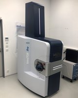
timsTOF SCP mass spectrometer (Bruker Daltonics)

The instrument provides extremely high speed and sensitivity to tackle proteomes of single cells or post translational modifications in a few cells.  The trapped ion mobility spectrometry (TIMS) device accumulates and concentrates ions of a given mass and mobility, enabling a unique increase in sensitivity and speed. Besides single cell proteomics, the new mass spectrometer allows high sensitive peptides and proteins identification and quantification. 

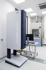
timsTOF Pro mass spectrometer (Bruker Daltonics)

timsTOF Pro mass spectrometer (Bruker Daltonics)

The timsTOF Pro mass spectrometer allows to provide the following services:


Peptide mass fingerprinting – identification of proteins from gel or solution including larger protein mixtures ; Characterization of posttranslational modification such as phosphorylation, glycosylation or disulphide bonds; Structural mass spectrometry: limited proteolysis, hydrogen/deuterium exchange, chemical cross-linking, covalent labelling; HPLC separation of peptides, proteins and small molecules (metabolites) coupled with mass spectrometric detection; Processing and interpretation of mass spectrometric data.

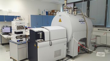
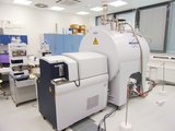
15T-SolariX XR FT-ICR mass spectrometer (Bruker Daltonics)

15T-SolariX XR FT-ICR mass spectrometer (Bruker Daltonics)

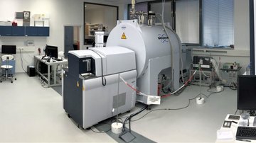
Bruker Daltonics 15T-Solarix XR FT-ICR mass spectrometer, with electrospray and MALDI ion sources. This ultra-high resolution mass spectrometer is used mostly for the determination of the precise mass of biological macromolecules, and the characterization of their posttranslational modifications. Further possibilities include peptide mass fingerprinting, detection of small molecule/metabolites, monitoring of protein structural changes and protein-protein interactions under physiological conditions by hydrogen-deuterium exchange, chemical cross-linking and covalent labelling.

Agilent Technologies 1200 HPLC system (usually coupled to the 15T-SolariX XR mass spectrometer) for the separation of complex peptide mixtures, proteins and metabolites.


This equipment allows to provide the following services:

  • Protein molecular weight determination by ultra-high resolution FT-ICR mass spectrometer with sequence confirmation by Top-down approach using different fragmentation techniques (collision induced dissociation, electron transfer/capture dissociation);
  • Peptide mass fingerprinting – identification of proteins from gel or solution including larger protein mixtures by using MALDI or ESI ;
  • Characterization of posttranslational modification such as phosphorylation, glycosylation or disulphide bonds;
  • Structural mass spectrometry: limited proteolysis, hydrogen/deuterium exchange, chemical cross-linking, covalent labelling;
  • HPLC separation of peptides, proteins and small molecules (metabolites) coupled with mass spectrometric detection by FT-ICR;
  • Processing and interpretation of mass spectrometric data.
MALDI-TOF (Bruker Daltonics)

MALDI-TOF (Bruker Daltonics)

Used for routine analysis of peptides and proteins.

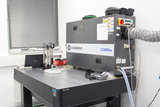
Excimer laser (Coherent)

Excimer laser (Coherent)

Used for Fast Photochemical Oxidation of Proteins (FPOP) - a type of hydroxyl-radical-based protein footprinting.

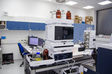
HPLC, UPLC (Agilent Technologies)

HPLC, UPLC (Agilent Technologies)

For the separation of complex peptide mixtures, proteins and metabolites.

Crystallization of proteins and nucleic acids

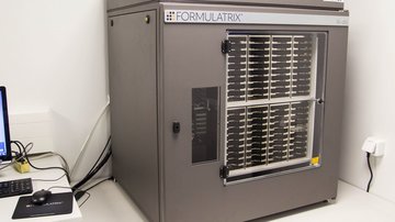
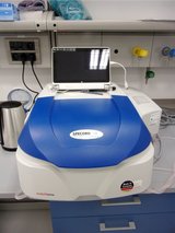
SONICC (Formulatrix)

SONICC (Formulatrix)

SONICC is using Second Harmonic Generation (SHG) and Ultraviolet Two-Photon Excited Fluorescence (UV-TPEF) in a completely automated imager to quickly image high throughput crystallization plates and positively identify protein crystals. SONICC is connected to the current RI1000 crystallisation hotel and is able to detect even extremely thin crystals, microcrystals <1 μm, and crystals obscured in birefringent LCP.

Vitrobot (Thermo Scientific)

Vitrobot (Thermo Scientific)

Equipment for preparation of samples suitable for electron diffraction and cryoEM experiments. Vitrobot from Thermo Scientific is able to vitrify samples on grids using liquid ethane. Instrumentation like this is crucial for the reproducibility of the procedure during the process of sample optimization.

SpectroLight 600 (Xtal Concepts)

SpectroLight 600 (Xtal Concepts)

SpectroLight 600 for measurement of dynamic light scattering in drops (including crystallisation experiments).

Detection of suitability of samples for crystallization or CryoEM grid preparation, together with in situ time lapse monitoring of protein crystallization and detection of nanocrystals for electron diffraction.

Crystallization hotel RI182 (Formulatrix)

Crystallization hotel RI182 (Formulatrix)

The hotel is able to store crystallization plates at 10 °C (SBS format, or Lipidic Cubic Phase) and image them according to the pre-set time schedule in visible light, polarized light, and UV light.

Dropsetter NT8 (Formulatrix)

Dropsetter NT8 (Formulatrix)

Automated pipetting crystallisation robot with controlled level of the air humidity, LCP head, possibility of robotic seeding.

Glovebox with stereomicroscope (GS)

Glovebox with stereomicroscope (GS)

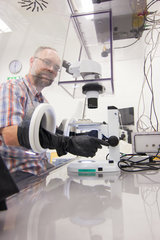
Glovebox with stereomicroscope (GS) for crystallization and crystal manipulation under defined (oxygen-free) atmosphere.

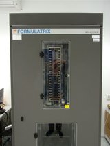
Crystallization hotel RI1000 (Formulatrix)

Crystallization hotel RI1000 (Formulatrix)

Crystallisation plate storage and automated crystallization monitoring enclosure allowing remote access to crystallization images. Crystallisation drop images can be taken using visible light, polarized light and UV; 500 positions for plates.

A service offered is the remote (web-based) access to crystallisation droplet images, and automated preliminary evaluation of crystallisation experiments.

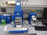
Gryphon Dropsetter (Art Robbins)

Gryphon Dropsetter (Art Robbins)

A multi-channel (96 channels) pipetting robot for the easy set-up of nanodrop crystallisation plates.

This crystallisation robot equipment allows the robotic setup of 96-well crystallisation plates, for screening of crystallisation conditions (and also for “routine crystal production”)  -  for proteins, nucleic acids, complexes of biological macromolecules.

Biophysical techniques

OCTET R8 (Sartorius)

OCTET R8 (Sartorius)

OCTET R8 (Sartorius) system for the kinetic and quantitative measurements using BioLayer Interferometry (BLI). It enables label-free analysis for the determination of kinetics and affinity of biomolecular binding based on BLI. Octet R8 is a fluidics-free, low maintenance detection system. Eight parallel, independent channels provide maximum speed, sensitivity and flexibility.

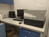
SPARK (TECAN)

SPARK (TECAN)

Spark microplate reader which offers absorbance (from 200 to 1 000 nm), fluorescence, including FRET and fluorescence polarization, and luminescence (ex 230 – 900 nm, em 280 – 900 nm) measurements. This technique provides a solution for ELISAs, low volume DNA/protein quantification and fast spectral scanning. It offers cuvettes and microplate formats up to 1 536 wells, has a higher performance, sensitivity and flexibility.

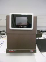
Surface Plasmon Resonance (SPR) system ProteOn XPR36 (BioRad)

Surface Plasmon Resonance (SPR) system ProteOn XPR36 (BioRad)

Label-free quantitative analysis of biomolecular interactions by the technique of surface plasmon resonance (SPR).

The ProteOnTM XPR36 protein interaction array system enables label-free quantitative analysis of biomolecular interactions in real time using SPR technology. The ProteOn system allows to screen analytes simultaneously against 36 different targets of interest, enabling rapid comparison among large numbers of interactions.


SPR can be used for:

  • Quantification of binding affinity and kinetics;
  • Determination of binding specificity and the number of binding sites;
  • Characterization of membranes, lipids, nucleic acids and micellar systems.


Sample requirements:

  • Concentration of ligand depends on the level of immobilization desired, generally 10–200 μg/ml. For kinetic analysis the best results are obtained by using a 100-fold range of analyte concentrations, 0.1–10xKd;
  • Immobilization of one interacting partner is essential. The service can provid with a sensor chip, or with the user bringing own chip;
  • The ProteOn acetate buffer (at pH 4.0, 4.5, 5.0, or 5.5) is recommended as immobilization buffer;
  • The recommended running buffer for most applications is the ProteOn phosphate buffered saline, pH 7.4 (10 mM sodium phosphate and 150 mM sodium chloride with 0.005% Tween 20).
Circular dichroism (CD) spectrometer Chirascan Plus (Applied Photophysics)

Circular dichroism (CD) spectrometer Chirascan Plus (Applied Photophysics)

Measurement of circular dichroism spectra and absorbance as function of temperature, pH and concentration to determine the secondary structure of proteins and peptides, conformation of RNA and DNA, as well as to detect conformational changes.

The Chirascan Plus CD spectropolarimeter with avalanche photodiode detector – provides fast scanning and high sensitivity. This instrument can simultaneously measure accurate CD, absorbance and fluorescence data.

Detection range: 170-1150 nm;

Peltier temperature control.


Circular dichroism can be used for:

  • Determination of protein folding;
  • Characterization of protein secondary structure and DNA conformation;
  • Detection of the changes in protein structure upon mutagenesis;
  • Studying of conformational stability of proteins and DNA (pH stability, denaturant stability, temperature, buffers addition of stabilizers).


Data processing:

The CDNN software package is available for detailed model-based analysis and predicting secondary structure using CD data;

Software Global Analysis of multiwavelength kinetic data is available to fit multi-dimensional experimental data to one of a number of specified models.


Sample requirements:

  • Measurement of CD spectrum for the determination of secondary structure of protein requires 160 µl of 0.1 – 0.2 mg/ml protein solution;
  • Measurement of CD spectrum for the determination of DNA conformation requires 160 µl of 20 µM of solution or 1400 µl of 2 µM solution;
  • Not optimal for CD solutions, containing DTT, imidazole, glycerol, DMSO, high concentrations of salts.
Monolith microscale thermophoresis (MST) NT.115 (Nano Temper)

Monolith microscale thermophoresis (MST) NT.115 (Nano Temper)

Used to study biomolecular interactions. The device allows to characterize protein-protein and protein-ligand (small molecule, DNA, RNA, peptides, sugars, lipids) interactions that can be measured under close to native conditions based on thermophoretic effect. Protein labeling is required with this device.

The Monolith NT.115 MST device allows to detect changes in hydration shell, charge or size of molecules and thus to detect biomolecular interactions.


MST can be used for:

Determination using a fluorescent dye or fluorescent protein of the affinity of interaction from 1nM to mM.


Sample requirements:

  • Concentration of fluorescent labeled molecule: 10 nM – 10 mM;
  • Final concentration of unlabeled molecule should be at least two orders of magnitude above the expected Kd value. To perform simulations of binding events and to help choose the appropriate concentration, the “Concentration Finder” software is available on the device control panel;
  • At least 20 µl samples per capillary is needed.
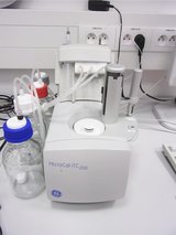
Isothermal titration calorimeter MicroCal iTC200 (Malvern Panalytical)

Isothermal titration calorimeter MicroCal iTC200 (Malvern Panalytical)

Label-free solution studies of biomolecular interactions.

The Malvern iTC200 instrument is used for the characterization of biomolecular interactions of small molecules, proteins, antibodies, nucleic acids, lipids etc.

 
The iTC200 device can be used for:

  • Direct measurement of submilimolar to nanomolar binding constants (10 3 – 10 9 M -1);
  • Thermodynamic characterization of the molecular interaction in a single experiment (stoichiometry, Kd, ∆H and ∆S values);
  • Calorimetric measurement over a range of biologically relevant conditions (temperature, salt, pH, etc.).
     

Sample requirements:

  • The buffer solution, containing both the macromolecule and the ligand of interest, should be the same.
  • The volume of the sample placed in the cell must be at least 300 µl. Preferably, the solutions of macromolecules should be dialysed against the buffer solution used for the ITC measurement;
  • The ligand solution (the sample placed in the injection syringe) must have a volume at least 70.0 µl.
  • Normally the ligand concentration should be 10 times as high as the concentration of macromolecule;
  • In the case of high affinity interactions, the minimum concentration of macromolecule (that causes measurable heat effects) is 10 µM. For low affinity interactions the macromolecule sample concentration should be at least 5 times the Kd value;
  • The buffers used should have low ionization enthalpies (e.g. phosphate, citrate, acetate);
  • If the presence of reducing agent is required for a protein stability, then ß‑mercaptoethanol (at a concentration lower than 5 mM) or TCEP (lower than 2 mM) should be used rather than DTT.
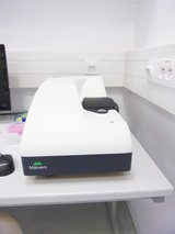
Dynamic light scattering (DLS) technique  Zetasizer Nano ZS90 (Malvern Panalytical)

Dynamic light scattering (DLS) technique  Zetasizer Nano ZS90 (Malvern Panalytical)

Measurement of molecular size using Dynamic Light Scattering (DLS), zeta potential and molecular weight using Static Light Scattering.

The Zetasizer Nano ZS90 instrument is used for the measurement of particle and molecular size using Dynamic Light Scattering, with the option of measuring zeta potential and electrophoretic mobility, and molecular weight using Static Light Scattering.

Size (diameter): from 0.3 nm to 5 microns;
Molecular weight measurement down to 10 kDa;
Temperature range 0-90°C.
 

Established methodologies and provided services:

  • Particle size analysis (hydrodynamic radius);
  • Temperature range 0-90°C.

Sample requirements:

  • 25 µl of sample and the same volume of “empty“ buffer ;
  • For protein solutions, concentrations of at least 0.2 mg/ml;
  • For the measurements of zeta potential in folded capillary cells, 0.75 ml of sample is required.
FTIR spectrometer Vertex 70v (Bruker)

FTIR spectrometer Vertex 70v (Bruker)

Provides the unique possibility to acquire a complete far and mid IR spectrum from 6000 cm-1 to 50 cm-1 in a single step measurement.

Multi-angle dynamic light scattering (MADLS) technique  Zetasizer Ultra (Malvern Panalytical)

Multi-angle dynamic light scattering (MADLS) technique Zetasizer Ultra (Malvern Panalytical)

Zetasizer Ultra is an instrument for the fast and accurate particle and molecule sizing, particle charge (zeta potential) and particle concentration measurements, and represents the most intelligent and flexible instrument in the Zetasizer range.

Most advanced system for the measurement of particle and molecular size, particle charge and particle concentration. The measurement of particle concentration is calibration-free and suitable for a wide range of materials.

It combines DLS and ELS system, incorporating Non-Invasive Back Scatter (NIBS) and Multi-Angle Dynamic Light Scattering (MADLS) technology for the measurement of particle and molecular size.

Monolith microscale label free thermophoresis NT.LabelFree (Nano Temper)

Monolith microscale label free thermophoresis NT.LabelFree (Nano Temper)

characterization of protein-ligand interactions based on thermophoretic effect, using the intrinsic tryptophan fluorescence. No sample modification is required with this device.

The NT.LabelFree MST instrument uses intrinsic tryptophan fluorescence for microscale thermophoresis detection, thereby allowing label-free and immobilization-free experiments.


MST can be used for:

The label and immobilization free determination of protein binding to Ions, nucleic acids, small molecules and sugars (with an affinity of interaction in the range of 10 nM to mM).


Sample requirements:

  • Concentration range of tryptophan-containing protein: 100 nM-10 µM;
  • Final concentration of unlabeled molecule should be at least an order of magnitude or more above the expected Kd value ;
  • Molecular weight range: 10-107 Da;
  • Minimum sample volume used: 10 µl per sample
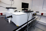
Modular fluorescence spectrometer FLS1000 (Edinburgh Instruments)

Modular fluorescence spectrometer FLS1000 (Edinburgh Instruments)

For measuring spectra from the ultraviolet to the mid-infrared spectral range, and lifetimes spanning from picoseconds to seconds.

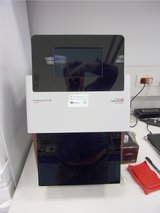
Differential scanning fluorescense (DSF) assay Prometheus NT.48 (Nano Temper)

Differential scanning fluorescense (DSF) assay Prometheus NT.48 (Nano Temper)

Measurement of protein stability using tyrosine and tryptophan fluorescence.

The Prometheus NT.48 instrument measure native DFS to determine protein thermal transition temperatures and stability of 48 up to samples at a time.

No dye is required, tryptophan fluorescence at 330 nm and 350 nm is detected;
Temperature range: from 15 °C to 95 °C.
 

DFS can be used for:

Determination of thermal transition temperatures and stability of proteins.
 

Sample requirements:

  • Protein must contain tryptophans in order to detect protein unfolding;
  • Sample concentration range: from 5 µg/ml to 250 mg/ml;
  • Prepare at least 20 µl of your samples;
  • For thermal unfolding experiments no assay development or special sample preparation is needed.
UV/Vis Spectrometer Specord 50 Plus  (Analytica Jena)

UV/Vis Spectrometer Specord 50 Plus (Analytica Jena)

Molecular absorption spectroscopy with ultraviolet and visible radiation in the spectral range 190 to 1100 nm.

The AnalyticJena SPECORD 50 PLUS device is a UV/Vis double-beam spectrophotometer with split-beam technology that combines high energy throughput with good stability.

  • 190-1100 nm;
  • 50-1500 µl of sample;
  • Scanning, dual beam
  • Temperature control with Peltier element, scan-range 5-95ºC.
     

The spectrometer can be used for:

  • Proteins and DNA thermostability measurements;
  • With or without stirrer can be used for enzyme kinetics.
Differential scanning calorimeter MicroCal VP-Capillary DSC (Malvern Panalytical)

Differential scanning calorimeter MicroCal VP-Capillary DSC (Malvern Panalytical)

Direct measurement of intramolecular stability of biological macromolecules, as well as the intermolecular stability of biologically-relevant complexes such as oligomeric proteins, nucleic acid duplexes, and micellar systems (lipid and detergent micelles).

The MicroCal VP-DSC instrument measures the temperature of thermally-induced structural transitions of molecules in solution. A complete thermodynamic profile is generated to understand the factors that affect conformation and stability of proteins, nucleic acids, micellar complexes and other macromolecular systems.

The operating temperature range is of -10°C to 130°C;
Maximum scan rates are 90°C/hr in the upscan mode and 60°C/hr in the downscan mode.
 

DSC can be used for:

  • The determination of transition midpoint, enthalpy (ΔH) of unfolding due to heat denaturation and change in heat capacity (ΔCp);
  • The study of factors that contribute to the folding and stability of native biomolecules, including hydrophobic interactions, hydrogen bonding, conformational entropy, and physical environment.


Sample requirements:

  • Sample buffer and buffer in the reference cell should be exactly the same;
  • The sample solutions should be dialysed against the buffer solution used for the DSC measurements.
  • Sample and reference cell volumes are 200 µl;
  • Typical sample concentration: 0.2 – 2.0 mg/ml;
  • If the presence of reducing agent is required for the sample, the use of up to 5 mM ß-mercaptoethanol or TCEP instead of DTT is recommended;
  • Since fluoride-containing samples cause irreparable damage to the VP-DSC cell, their use is prohibited.

Diffraction techniques

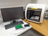
SAXSpoint 2.0 (Anton Paar)

SAXSpoint 2.0 (Anton Paar)

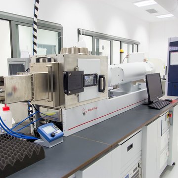
Versatile instrument to perform SAXS or WAXS experiments on liquid samples, in the temperature range -10° C to 120° C. The Small Angle X-ray Scattering instrument is equipped with the latest high brilliance source MetalJet C2+ having a liquid gallium alloy anode, and a Dectris EIGER 1M detector. Samples can be loaded using the automated high precision sampler or manually in capillaries, including a high S/N silicon nitride measurement cell. The state of the sample can be monitored online using UV-Vis spectroscopy to identify radiation damage, measure precise sample concentration and aggregation state.
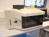
D8 Venture diffractometer (Bruker)

D8 Venture diffractometer (Bruker)

Bruker D8 Venture diffractometer with a high-flux liquid Gallium X-ray source MetalJet D2, Photon II detector and Kappa goniometer. The diffractometer is used (at CMS) for X-ray diffraction of biomolecular crystals.
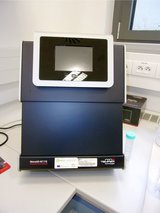
ISX stage for D8 Venture – motorized stage for in-situ X-ray diffraction experiments, enabling screening of diffraction properties in crystallization trays.


This equipment allows to offer the following services:

  • In-situ (in the crystallisation plates) testing of crystal diffraction using the ISX stage;
  • Testing of diffraction using mounted crystals and / or measurement of X-ray diffraction data;
  • Diffraction data processing, providing a data file (such as an MTZ file);
  • Assistance / advice to solve a 3D structure (including a full 3D structure determination service on request);
  • Upon request: Measurement of X-ray diffraction data sets at synchrotron radiation sources.

Team

Ing. Jan Dohnálek, Ph.D. Ing. Jan Dohnálek, Ph.D.

Ing.
Jan Dohnálek, Ph.D.

Head of the CMS Core Facility, Biocev representative of Czech infrastructure for integrative structural biology - CIISB, Biocev representative of European infrastructure Instruct

jan.dohnalek@ibt.cas.cz
+420325873758
Tatsiana Charnavets, Ph.D. Tatsiana Charnavets, Ph.D.

Tatsiana Charnavets, Ph.D.

Scientist in charge of the biophysical measurements service

tatsiana.charnavets@ibt.cas.cz
+420325873789
RNDr. Jiří Pavlíček, Ph.D. RNDr. Jiří Pavlíček, Ph.D.

RNDr.
Jiří Pavlíček, Ph.D.

Scientist in charge of the crystallization and X-ray diffraction service (including SAXS)

jiri.pavlicek@ibt.cas.cz
+420325873787
Ing. Jan Stránský, Ph.D. Ing. Jan Stránský, Ph.D.

Ing.
Jan Stránský, Ph.D.

Scientist in charge of crystallisation and X-ray diffraction service (including SAXS)

jan.stransky@ibt.cas.cz
+420325873788
RNDr. Petr Pompach, Ph.D. RNDr. Petr Pompach, Ph.D.

RNDr.
Petr Pompach, Ph.D.

Scientist in charge of the mass spectroscopy service

petr.pompach@ibt.cas.cz
+420325873785
RNDr. Ľubica Škultétyová, PhD. RNDr. Ľubica Škultétyová, PhD.

RNDr.
Ľubica Škultétyová, PhD.

Research Assistant

lubica.skultetyova@ibt.cas.cz
+420325873780
Bc. Michal Strnad Bc. Michal Strnad

Bc.
Michal Strnad

computer maintenance and IT

michal.strnad@ibt.cas.cz
+420325873759
Mgr. Pavla Vaňková, Ph.D. Mgr. Pavla Vaňková, Ph.D.

Mgr.
Pavla Vaňková, Ph.D.

Mass Spectrometry

pavla.vankova@ibt.cas.cz
+420325873785