Laboratory of Transgenic Models of Diseases
A link between autoimmune disorders and tooth enamel defects revealed
In a groundbreaking study published in Nature, an international team of researchers has discovered a crucial link between autoimmune disorders and defects in tooth enamel development. This study sheds light on the poorly understood conditions of Amelogenesis Imperfecta in patients with Autoimmune Polyglandular Syndrome Type-1 (APS-1) and Celiac Disease.
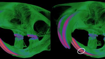
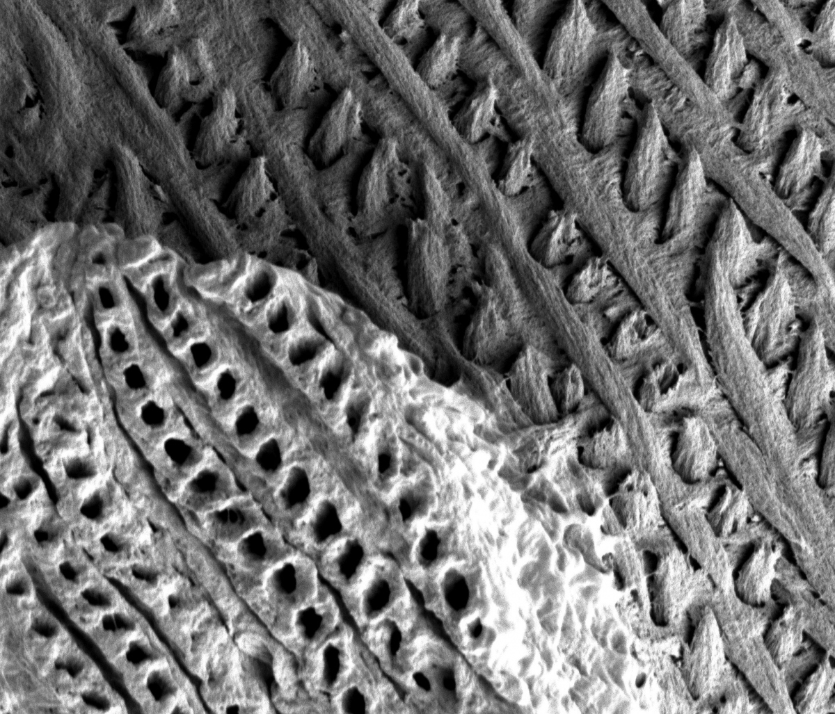
Ameloblasts, the cells responsible for tooth enamel formation, rely on several proteins to create a durable outer layer for our teeth. When these proteins are attacked by the body’s immune system, it leads to Amelogenesis Imperfecta, a condition characterized by weakened, discolored, and easily damaged teeth. This condition has long puzzled the medical community, but the new study led by Jakub Abramson from the Weizmann Institute in Israel offers unprecedented insights.
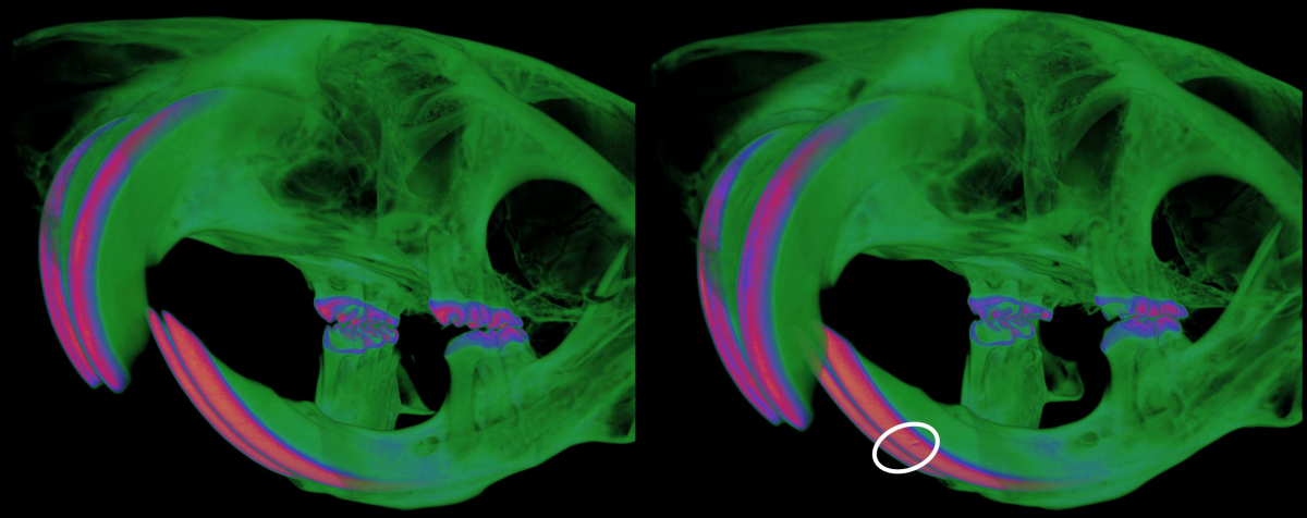
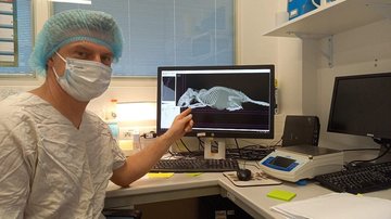
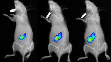
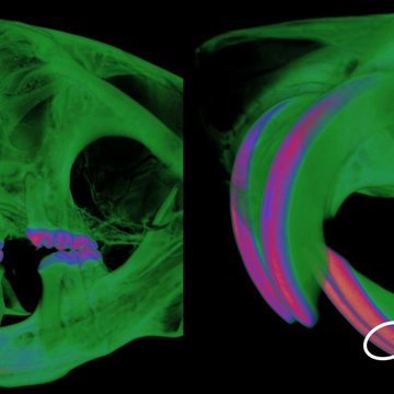
The research team, including notable contributions from Dr. Jan Prochazka from Czech Center for Phenogenomics, has utilized advanced technology to analyze tooth phenotypes, shedding light on the processes disrupting enamel mineralization in autoimmune conditions. The study reveals that most patients with APS-1 and Celiac Disease develop autoantibodies, particularly of the IgA isotype, against ameloblast-specific proteins. This results in a breakdown of the body’s tolerance to these proteins, leading to compromised enamel formation. In Celiac Disease, this phenomenon appears to be linked to a loss of tolerance to intestinal antigens that are also present in enamel tissue. These findings suggest a novel type of IgA-dependent autoimmune disorder, which the researchers have collectively termed “autoimmune amelogenesis imperfecta.”
Publication:
Gruper, Y., Wolff, A. S. B., Glanz, L. et al. Autoimmune amelogenesis imperfecta in patients with APS-1 and coeliac disease. Nature (2023). https://doi.org/10.1038/s41586-023-06776-0 / https://www.nature.com/articles/s41586-023-06776-0
Photo: Weizmann Institute of Science